Anatomy
Anatomy is a branch of biology that deals with the structure of plants and animals. Comparative anatomy is a related field in which the structures of different animals are studied and compared. There are three main areas of anatomy: gross anatomy deals with organs and organ groupings called systems that are visible to the naked eye; cytology is the study of cell structure; and histology examines the structure of tissues. Microscopes are used in both cytology and histology to study cell and tissue structures.
History of anatomy
Attempts to understand the structure of living things go as far back as Aristotle (384–322 B.C. ), the famous Greek philosopher and biologist. His dissection (cutting into pieces to examine the parts) and study of animals and plants led to his formation of a classification system that was used by scientists for almost 2,000 years.
Some of the first human dissections were carried out by Greek anatomists and physicians Herophilus (late fourth century B.C. ) and his younger follower Erasistratus. Herophilus made many anatomical studies of the brain. He distinguished the cerebrum (larger portion) from the cerebellum (smaller portion), suggested that the brain was the seat of intelligence, and identified and named several structures of the brain, some of which still carry the names he gave them. He also discovered that nerves originate in the brain and noted the difference between motor nerves (those concerned with motion) and sensory nerves (those related to sensation). Together with Erasistratus, Herophilus established the disciplines of anatomy and physiology (the science that deals with the function of the body's parts and organs).
In his studies of the heart and blood vessels, Erasistratus came very close to working out the circulatory system of the blood. He understood that the heart served as a pump and he studied and explained the function of the heart valves. Erasistratus theorized that the arteries and veins both spread from the heart but incorrectly believed that the arteries carried air instead of blood.
After Erasistratus's time, the dissection of human bodies to study their anatomy ended due to the pressure of public opinion. Egyptians believed that a body needed to remain whole to enter the afterlife, and they engaged in the practice of mummification (treating a body with preservatives for burial).
Important contributions to the science of anatomy were made by the last and most influential of the great ancient medical practitioners, Greek physician Claudius Galen ( A.D. 131–200). He expertly dissected and accurately observed all kinds of animals but sometimes mistakenly applied what he saw to the human body. Nevertheless, he was the first to observe that muscles work in opposing pairs: for every muscle that causes a joint to bend, there is an opposing muscle that restores the joint to its original position.
Through experiments, Galen observed and described two ground-breaking anatomical events: (1) paralysis resulting from the cutting of the spinal cord and (2) the process by which urine passes from the kidneys to the bladder. In his observations about the heart and blood vessels, however, Galen made critical errors that remained virtually unchallenged for 1,400 years. He mistakenly believed that blood was formed in the liver and was circulated throughout the body by the veins. When anatomical research stopped for many centuries, Galen's teachings remained the ultimate medical authority.
After human dissections resumed in the sixteenth century, the long-held teachings of Galen were overturned by the work of Flemish anatomist and physician Andreas Vesalius (1514–1564). Vesalius, who founded modern scientific anatomy, noted obvious conflicts between what he saw in his dissections of the human body and what Galen had described. He reasoned that Galen's errors resulted from only having done animal dissections, which often did not apply to human anatomy.
In 1543, Vesalius published one of the most important books in medical history and the world's first textbook of anatomy, On the Structure of the Human Body. The book contains detailed anatomical descriptions of all parts of the human body, directions for carrying out dissections, and meticulously drawn illustrations. Vesalius believed that accurate, basic knowledge of the human body could only be gained by performing human dissections. In his book, he set forth an objective, scientific method of conducting medical research that was to become the foundation of anatomical research and education throughout the world.
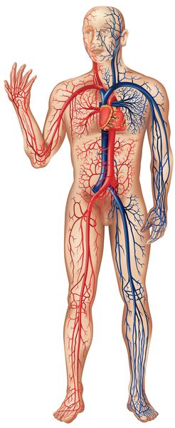
The correct description of the circulation of blood was provided by English physician William Harvey (1578–1657). In the course of many experimental dissections, he established the existence of pulmonary circulation (blood flowing from heart to lungs to heart) and noted the one-way flow of blood. He was the first to discover that blood flows in a continuous circle from the heart to the arteries to the veins and back to the heart. Harvey published this radical new concept of blood circulation in 1628.
The discovery of capillaries (small blood vessels) by Italian anatomist Marcello Malpighi (1628–1694) in 1661 provided the factual evidence to confirm Harvey's theory of blood circulation. Malpighi discovered the capillaries—the tiny connecting links between the veins and arteries—using the newly invented microscope.
The science of anatomy was further advanced by the work of English physicist Robert Hooke (1635–1703). His 1665 publication Micrographia describes the structures of insects, fossils, and plants in detail from his microscopic studies. While examining the porous structure of cork, Hooke coined the term "cells" to describe the tiny rectangular holes he observed. This led scientists to adopt the concept of cells as the unit
structures of tissues, which in turn led to the suggestion of cells as the building blocks of organs and to the discovery of the cell nucleus. A later theory proposing that all of the body's tissues are composed of cells was the basis for the science of cytology.
Histology, or the study of tissues (structured groups of specialized cells), began in earnest in the 1700s with the work of French scientist Xavier Bichat (1771–1802). Bichat found that organs were built up out of different types of simpler structures, and each of these simpler structures could occur in more than one organ. He further noted that different tissues have specific properties and are thereby vulnerable to tissue-specific diseases.
Until that time, general anatomy was a descriptive order, based upon obvious characteristics such as the location of organs. Bichat suggested adopting a systematic order for anatomy based upon structure and function. He specified 21 tissues (or systems) in the human body based on what he saw with his naked eye, distinguishing these different tissues by their composition and by the arrangement of their fibers. These include epithelial (skin and digestive), muscular, nervous, connective, and vascular (blood) types.
Histology began to take on its modern form with the introduction of cell theory in 1839. At that time tissues began to be understood not as the basic building blocks of living things but as unique systems of cells with their own stages of development within the embryo (early stage of an organism's growth before birth or hatching).
Modern anatomy
Anatomy today makes use of knowledge from many fields of science to explore and understand how the structure of an organism's cells, tissues, and organs relates to their function.
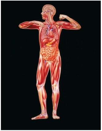
Human anatomy, a crucial element in the medical school curriculum, divides the body into separate functional systems. These consist of the skin, the muscles, the skeleton, the circulatory system (blood, blood vessels, and heart), the digestive system, the urinary system, the respiratory system (lungs and breathing), the nervous system (brain, spinal cord, and nerves), the endocrine system (glands and hormones), and the reproductive system.
[ See also Circulatory system ; Digestive system ; Endocrine system ; Muscular system ; Nervous system ; Reproductive system ; Respiratory system ; Skeletal system ]